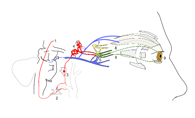
Scheme showing sympathetic and parasympathetic innervation of the pupil and sites of lesion in a Horner's syndrome.
1. sympathetic fibers arise from the hypothalamus
2. stellate ganglion
3. synapse at the superior cervical ganglion
4. sympathetic plexus around internal carotid artery
5. oculomotor nerve (Cranial nerve 3) fibers synapse at the ciliary ganglion (blue)
6. Short ciliary nerves from ciliary ganglion carrying parasympathetic supply to sphincter pupillae (green)
7. Trigeminal fibers (Cranial nerve 5) relay in ciliary ganglion and carry sympathetic supply (yellow)
8. Long ciliary nerve fibers (from the ophthalmic branch of CN 5) are the afferent limb of the blink reflex carrying sensory information from the cornea.
9. Sphincter pupillae (circular fibers) and Dilator pupillae (radial fibers) muscles of the pupil.
Near the stellate ganglion, the sympathetic fibers go around the sublavian artery (shown along with the carotid vessels). This is a site of lesion especially due to its proximity to the apex of the lung (eg. Pancoast's tumor). The superior division of oculomotor nerve is shown supplying the Superior rectus and levator palpebrae superioris.
commons.wikimedia.org/wiki/File:Horner%27s_Syndrome_and_Autonomic_innervation_of_the_eye.svg